Scientists showed that they could alter brain activity of rats and either wake them up or put them in an unconscious state by changing the firing rates of neurons in the central thalamus, a region known to regulate arousal. The study, published in eLIFE, was partially funded by the National Institutes of Health.
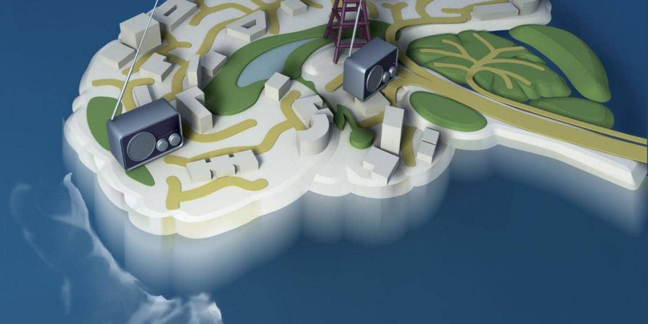
“Our results suggest the central thalamus works like a radio dial that tunes the brain to different states of activity and arousal,” says Jin Hyung Lee, PhD, assistant professor of neurology, neurosurgery and bioengineering at Stanford University, and a senior author of the study, in a release.
Located deep inside the brain, the thalamus acts as a relay station sending neural signals from the body to the cortex. Damage to neurons in the central part of the thalamus may lead to problems with sleep, attention, and memory. Previous studies suggested that stimulation of thalamic neurons may awaken patients who have suffered a traumatic brain injury from minimally conscious states.
Lee’s team flashed laser pulses onto light sensitive central thalamic neurons of sleeping rats, which caused the cells to fire. High frequency stimulation of 40 or 100 pulses per second woke the rats. In contrast, low frequency stimulation of 10 pulses per second sent the rats into a state reminiscent of absence seizures that caused them to stiffen and stare before returning to sleep.
“This study takes a big step towards understanding the brain circuitry that controls sleep and arousal,” says Yejun (Janet) He, PhD, program director at NIH’s National Institute of Neurological Disorders and Stroke (NINDS).
When the scientists used functional magnetic resonance imaging (fMRI) to scan brain activity, they saw that high and low frequency stimulation put the rats in completely different states of activity. Cortical brain areas where activity was elevated during high frequency stimulation became inhibited with low frequency stimulation. Electrical recordings confirmed the results. Neurons in the somatosensory cortex fired more during high frequency stimulation of the central thalamus and less during low frequency stimulation.
“Dr Lee’s innovative work demonstrates the power of using imaging technologies to study the brain at work,” says Guoying Liu, PhD, a program director at the NIH’s National Institute of Biomedical Imaging and Bioengineering (NIBIB).
How can changing the firing rate of the same neurons in one region lead to different effects on the rest of the brain?
Further experiments suggested the different effects may be due to a unique firing pattern by inhibitory neurons in a neighboring brain region, the zona incerta, during low frequency stimulation. Cells in this brain region have been shown to send inhibitory signals to cells in the sensory cortex.
Electrical recordings showed that during low frequency stimulation of the central thalamus, zona incerta neurons fired in a spindle pattern that often occurs during sleep. In contrast, sleep spindles did not occur during high frequency stimulation. Moreover, when the scientists blocked the firing of the zona incerta neurons during low frequency stimulation of the central thalamus, the average activity of sensory cortex cells increased.
Although deep brain stimulation of the thalamus has shown promise as a treatment for traumatic brain injury, patients who have decreased levels of consciousness show slow progress through these treatments.
“We showed how the circuits of the brain can regulate arousal states,” says Lee. “We hope to use this knowledge to develop better treatments for brain injuries and other neurological disorders.”
Image courtesy of courtesy of Lee lab, Stanford University, CA


