Orexin is important in regulating the switch between sleep and wakefulness in the brain. If a person lacks adequate orexin neurons, then they have narcolepsy, where sleep and wakefulness are inadequately switched on and off.
When people with narcolepsy exhibit cataplexy, muscle atonia that can be triggered by strong emotion, that is theorized to be the muscle paralysis of REM sleep taking place while a person is awake. An international research team led by Kanazawa University previously found two types of neurons preventing narcolepsy by receiving orexin from orexin neurons: one is noradrenaline neurons in the locus coeruleus of the brain, suppressing strong sleepiness; and the other is serotonin neurons in the dorsal raphe nucleus of the brain, inhibiting cataplexy.
In a new study published in PNAS [Proceedings of the National Academy of Sciences], the research team has discovered that serotonin neurons in the dorsal raphe nucleus inhibit catalepsy by reducing activities of the amygdala, a brain region essential in processing emotional responses as well as in emotional memory.
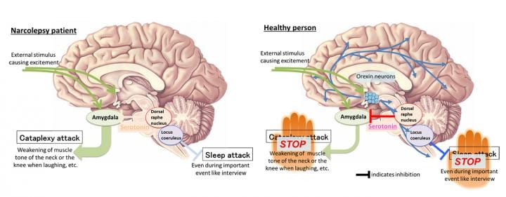
External stimulus causing excitement such as laughter by a joke augments the amygdala activity. In a narcolepsy patient (left) lacking orexin neurons, activities of the amygdala become excessive, causing cataplexy. In healthy person (right), orexin neurons augment the activities of serotonin neurons in the dorsal raphe nucleus, which reduce activities of the amygdala due to increased release of serotonin in the amygdala, which in turn inhibits cataplexy. Credit: Kanazawa University
Serotonin neurons in the dorsal raphe nucleus extend projections throughout the brain and send information. In this study, with an optogenetic tool, the team discovered that catalepsy was almost completely inhibited by artificial augmentation of serotonin release; the release was induced by selectively stimulating serotonin nerve terminals in the amygdala in narcolepsy model mice.
In addition, the team found that serotonin release reduced amygdala activity. When the amygdala activity was artificially reduced in a direct manner, cataplexy was inhibited; while artificially augmented, frequency of cataplexy attacks increased. Furthermore, the effect of orexin neurons inhibiting cataplexy was found to be abolished when serotonin release was inhibited selectively in the amygdala.
The same experimental operation in the brain region that controls REM sleep did not inhibit cataplexy.
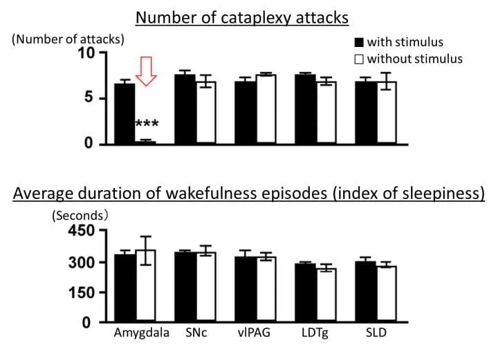
When serotonin nerve terminals in the amygdala are stimulated to augment serotonin release, cataplexy is inhibited (indicated by arrow in the upper part) while sleepiness is not inhibited (lower part). On the other hand, stimulation of serotonin nerve terminals in the other region controlling REM sleep does not induce any changes. LDTg, laterodorsal tegmental nucleus; SLD, sublaterodorsal nucleus; SNc, substantia nigra compact part; vlPAG, ventrolateral periaqueductal gray. Credit: Kanazawa University
This study reveals that serotonin neurons do not directly suppress muscle tone weakening but inhibit cataplexy by reducing and controlling activities of the amygdala, which is involved in communicating emotional excitement. In fact, it is known that the amygdala of narcolepsy patients without orexin neurons responds excessively when the patients see, for example, interesting photos.
By identifying this neuronal pathway, the team states that the current study has made a leap forward in understanding of the whole picture of narcolepsy’s mechanism. They also think this discovery may ultimately guide the development of new therapies for cataplexy.


