

Excessive fragmentary myoclonus (EFM), previously called excessive fragmentary hypnic myoclonus, is defined by the American Academy of Sleep Medicine (AASM) as at least five characteristic fragmentary myoclonus electromyographic potentials per minute during at least 20 minutes of NREM sleep. EFM is scored using an optional rule (VII Movement Rules, Section 4) in the AASM scoring manual (Table 1).1 These electromyographic potentials are usually seen as muscle surface potentials of the tibialis anterior muscle with an amplitude of 50 to 200 microvolts and a duration of less than 150 milliseconds.2,3 Patients may describe these feelings as twitches or cramps, or they may be oblivious to any physical discomfort.
There are few published reports on the topic. In 1984, Broughton and colleagues first described this polysomnographic finding in a patient with excessive daytime somnolence.2 The clinical significance of EFM is not clear, but the polysomnographic findings are interesting, and the diagnosis can be overlooked since the muscle groups involved are small and do not usually lead to visible limb motion. The case of patient Mr ZZ provides a basis for discussion of polysomnographic findings, manifestations, and current thoughts on treatment of EFM.
PATIENT CASE
Mr ZZ is a 78-year-old male (BMI 27) who presented for the evaluation of restless sleep. He had been having symptoms for several years, but due to a busy lifestyle, he had no prior sleep evaluation. He was reported to have snoring events, and he complained of a long sleep latency period at home. The patient discussed having persistent leg twitches and cramps throughout the night, which disturbed his sleep and made him feel poorly rested in the morning; he denied symptoms consistent with a diagnosis of restless leg syndrome (RLS) such as improvement with movement and abnormal sensations in legs.
He did not report significant sleepiness; Epworth Sleepiness Scale score was 1. The patient did not have any ancillary symptoms associated with narcolepsy such as cataplexy, hypnagogia, or sleep paralysis.
Significant comorbid factors included diabetes and back pain. Current medications reported by the patient included amlodipine, verapamil, lisinopril, bumetanide, allopurinol, atorvastatin, pioglitazone, sitagliptin, Metanx (multivitamin), vitamin D, Ecotrin (aspirin), and Colace (stool softener).
POLYSOMNOGRAPHY
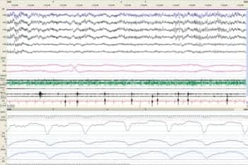
Figure 1. REM stage sleep, the characteristic EMG findings of excessive fragmentary myoclonus are marked with black arrows.
He underwent a standard overnight polysomnogram, which showed an apnea/hypopnea index (AHI) of 8.5 events per hour with a REM AHI of 21.9 events/hour and supine AHI of 58.1 events/hour, a respiratory disturbance index (RDI) of 25.4 events/hour of sleep, and an oxygen nadir of 83%. The oxygen saturation was persistently 88% to 90% during sleep even when not associated with apneas or hypopneas. There were no significant limb movements noted on the video monitoring.
On review of his polysomnogram, frequent, small (<150 milliseconds), repetitive, alternating muscle twitches were noted during both REM (Figure 1) and non-REM sleep (Figure 2).
According to the AASM, EFM may be a benign movement phenomenon associated with a characteristic EMG pattern. In many cases, no visible movements are present. Gross jerk-like movements across the joint spaces are not observed. When minor movement across a joint space is present, the movement resembles the small twitch-like movements of the fingers, toes, and the corner of the mouth that are intermittently seen during REM sleep in normal individuals.1 In comparison, nocturnal leg cramps are a clinical complaint without EMG findings and periodic limb movements are a series of at least four limb movements lasting 0.5 to 10 seconds that are at least 8 microvolts above the baseline.

Figure 2. Non-REM stage sleep, the characteristic EMG findings of excessive fragmentary myoclonus are marked with black arrows.
DIAGNOSIS
Mr ZZ was given the diagnosis of obstructive sleep apnea (OSA) syndrome and excessive fragmentary myoclonus.
TREATMENT
He was prescribed an autotitrating positive airway pressure (APAP) machine set at 7 to 12 cm H2O based on a PAP titration study showing benefit at these pressures varying by body position. He was very compliant with the APAP device but felt that it did not improve his sleep quality or duration. His twitching and cramping symptoms were distressing, and he was offered a dopamine agonist as a trial of therapy.
This patient and his subsequent course are typical for EFM patients. They are usually older men with other sleep-related disorders who are found to have persistent hypoxia on polysomogram.4 His course of treatment also follows that of other EFM patients we have encountered. He had OSA, which was treated, but his leg twitching and cramping did not resolve nor did his restless sleep.
In cases like this one, the clinician is faced with a dearth of objective evidence for effective treatments because the literature infers that this is not a significant cause of distress or symptoms for patients. In our institution, other comorbid causes of restless sleep (OSA, insomnia, medications) are addressed first. We measure the patient’s thyroid, vitamin and iron stores, and electrolytes similar to the workup we perform for RLS. If no other cause is found and the EFM symptoms persist, we offer patients a dopamine agonist trial for a fixed period of time to look for clinical efficacy. We occasionally offer patients quinine, after explaining that the Food and Drug Administration (FDA) has declared that the medication should not be used for nighttime leg cramps. We also explain that the FDA has placed a black-box warning regarding severe bleeding problems, kidney damage, irregular heartbeat, and severe allergic reactions with the use of quinine for leg cramps and allow the patient to make an educated decision using a risks/benefits analysis.
DISCUSSION
The origin of EFM has not been clearly delineated. Previous studies in animals suggested that myoclonic twitches are caused by phasic reticulospinal volleys descending upon alpha motor neurons during sleep.5 More current authors suggest the brainstem6 or aberrant solitary motor neurons without higher level control4 as the origin for these twitches. The typical EMG activity can be seen in sleep and relaxed wakefulness and is a normal phenomenon in REM sleep in healthy individuals.
Movement Rules1
Scoring Rules for Excessive Fragmentary Myoclonus (Optional)
- The following rules define EFM:
- The usual maximum EMG burst duration seen in fragmentary myoclonus is 150 msec.
- At least 20 minutes of NREM sleep with EFM must be recorded.
- At least 5 EMG potentials per minute must be recorded.
Notes:
- EFM may be a benign movement phenomenon associated with a characteristic EMG pattern as there have been no reported clinical consequences.
- In many cases no visible movements are present. Gross jerk-like movements across the joint spaces are not observed. When minor movement across a joint space is present, the movement resembles the small twitch-like movements of the fingers, toes, and the corner of the mouth intermittently seen in REM sleep in normal individuals.
- In some cases when visible movement is present, the EMG burst duration may be >150 msec.
Severity can be assessed by calculating the fragmentary myoclonus index, as defined by Lins et al,3 as follows: Each 30-second scoring epoch is divided into ten 3-second mini epochs. The number of these mini epochs with at least one fragmentary myoclonus potential (fulfilling the criteria) is counted for each 30-second epoch, resulting in a number between 0 and 10 for each epoch. These events per epoch are totaled and divided by the number of hours of sleep. This method can be time-consuming and is not likely to be readily performed outside a research study.
When does the clinician need to intervene? The largest studies of EFM patients included 68 patients with mild to severe EFM, based on their fragmentary myoclonus index ranging from 4.0 to 370.9; it showed that the highest indexes were associated with male sex, elevated BMI, increasing age, and oxygen desaturation, and EFM was more often found in patients with a comorbid sleeping disorder.4 No clinical studies to date address EFM as a cause of clinical symptoms.
EFM can be overlooked as a diagnosis because the myoclonic EMG findings are normal in phasic REM sleep7,8 and the clinician must carefully observe non-REM periods to discover the diagnostic EMG potentials. Further complicating the diagnosis is the use of limb movement auto-scoring and filters, which may remove the classic pattern from the view of the clinician.
Excessive fragmentary myoclonus is a diagnosis of uncertain clinical importance, but it is often found in relation to other sleep disorders and the polysomnographic findings are striking. It is also often associated with nocturnal desaturations,4 advanced age, and male sex. There are no current recommendations for treatment other than expert opinion because many in the sleep field believe it to be clinically insignificant. In this case, the patient presented with what we felt were important clinical findings: restless sleep, desaturations noted on polysomnogram, and muscle cramping sensations while trying to sleep.
Garrett R. Bird, MD, CM; Vishal Sawhney, MD; Philip Alapat, MD; and Amir Sharafkhaneh, MD, practice at Baylor College of Medicine, Houston. They can be reached at [email protected].
REFERENCES
- American Academy of Sleep Medicine. International Classification of Sleep Disorders, Diagnostic and Coding Manual. 2nd ed. Westchester, Ill: American Academy of Sleep Medicine; 2005.
- Broughton R, Tolentino MA. Fragmentary pathological myoclonus in NREM sleep. Electroencephalogr Clin Neurophysiol. 1984;57(4):303-9.
- Lins O, Castonguay M, Dunham W, Nevsimalova S, Broughton R. Excessive fragmentary myoclonus: time of night and sleep stage distributions. Can J Neurol Sci. 1993;20(2):142-6.
- Frauscher B, Kunz A, Brandauer E, Ulmer H, Poewe W, Hogl B. Fragmentary myoclonus in sleep revisited: a polysomnographic study in 62 patients. Sleep Med. 2011;12(4):410-5.
- Gassel MM, Marchiafava PL, Pompeiano O. Phasic changes in muscular activity during desynchronized sleep in unrestrained cats. An analysis of the pattern and organization of myoclonic twitches. Arch Ital Biol. 1964;102:449-70.
- Vetrugno R, Plazzi G, Provini F, Liguori R, Lugaresi E, Montagna P. Excessive fragmentary hypnic myoclonus: clinical and neurophysiological findings. Sleep Med. 2002;3(1):73-6.
- Montagna P, Liguori R, Zucconi M, et al. Physiological hypnic myoclonus. Electroencephalogr Clin Neurophysiol. 1988;70(2):172-6.
- Dagnino N, Loeb C, Massazza G, Sacco G. Hypnic physiological myoclonias in man: an EEG-EMG study in normals and neurological patients. Eur Neurol. 1969;2(1):47-58.


