
Peripheral arterial tonometry is a physiologic sensing approach used in sleep medicine to infer respiratory disturbances by tracking autonomic nervous system activity during sleep. While early and widely used implementations relied on finger-based sensors, peripheral arterial tone signals can be measured at multiple body sites where arteries lie close to the skin surface. As the technologies have matured, they have become an important category within home sleep testing (HST), offering an easy-to-use alternative to both in-lab testing and HSTs that require more sensor connections.
During stable sleep, peripheral arterial tone remains relatively steady. But when a respiratory disturbance occurs, the stress activates the sympathetic nervous system, producing a rapid vasoconstrictive response in peripheral arteries. Peripheral arterial tone-based devices detect the change in volume in peripheral arteries that results from pauses in blood flow.
Peripheral arterial tone-based home sleep testing systems frequently also integrate complementary physiologic signals, such as heart rate dynamics and oxygen saturation. Typically, when arterial tone attenuation occurs in concert with other changes (such as heart rate acceleration and oxygen desaturation patterns) during sleep, algorithms can identify events that correspond to respiratory disturbances and arousals.
According to an American Academy of Sleep Medicine guideline published in 2017, peripheral arterial tonometry used in combination with oximetry and actigraphy is “technically adequate” to diagnose obstructive sleep apnea in patients likely to have the condition who do not have specific comorbidities.
Sleep Review‘s guide to peripheral arterial tone systems compares options on parameters including form factor, wavelengths, sampling rates, optical architecture, and more.
The systems compared are:
Compumedics Somfit/Somfit-D

EnsoData EnsoHST
SleepImage System

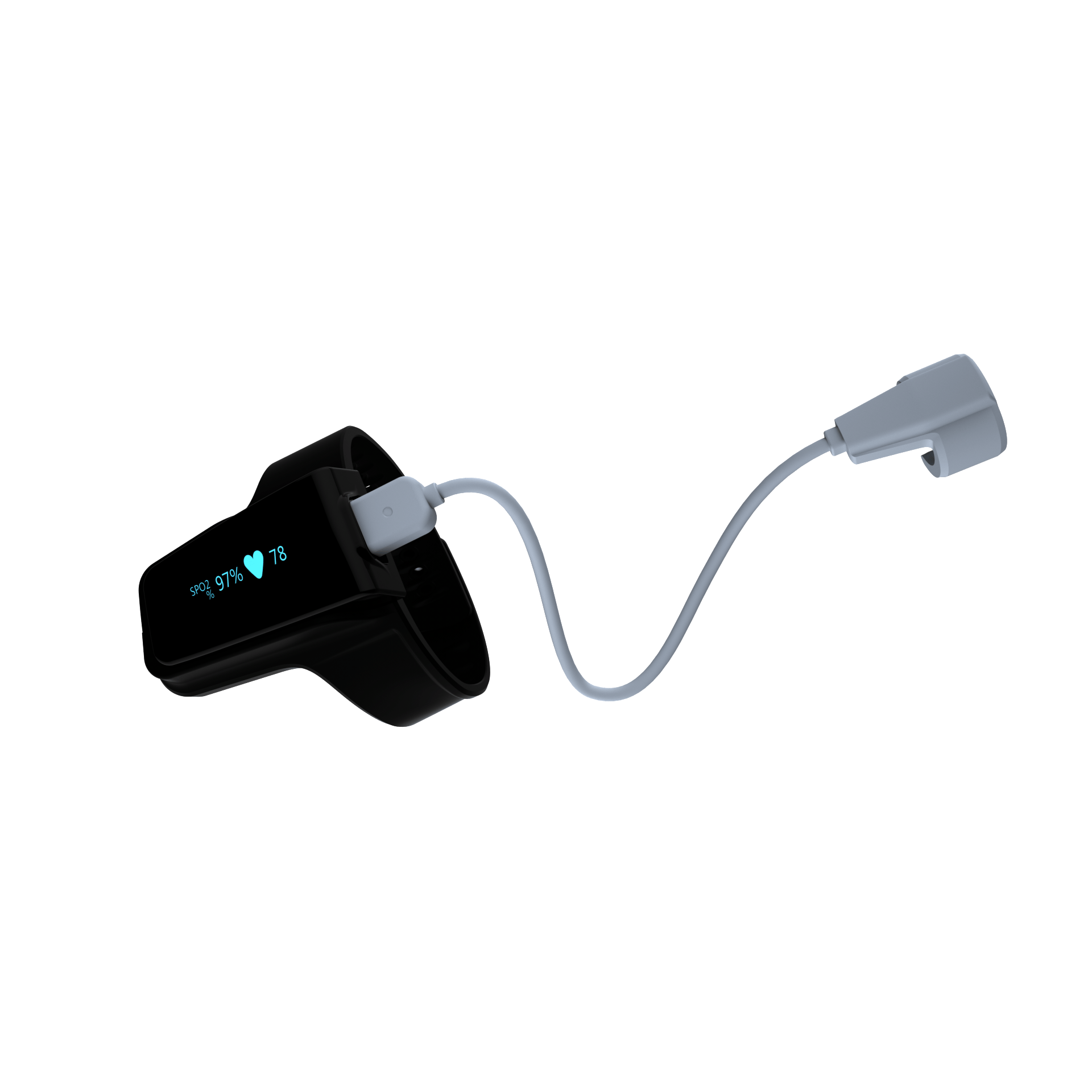
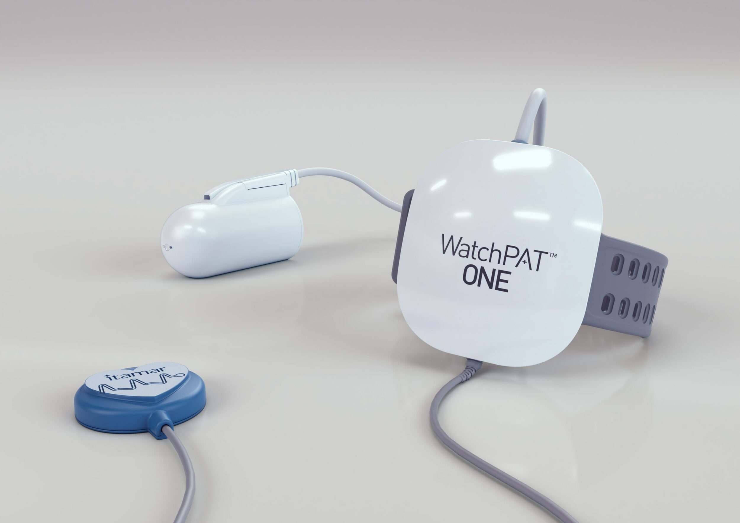
ZOLL Itamar WatchPAT ONE/WatchPAT 300
| Company: | Compumedics | EnsoData | SleepImage | ZOLL Itamar |
| Device: | Somfit/Somfit-D | EnsoHST | SleepImage System | WatchPAT ONE/WatchPAT 300 |
| Website: | compumedics.com | ensodata.com/ensohst | sleepimage.com | itamar-medical.com |
| Description: | The Somfit platform uses reflectance forehead oximetry at 0.1% resolution sampling at 78 Hz to return SPO2 and peripheral arterial tone—an optimal place due to the presence of 3 large peripheral arteries (supratrochlear artery, supraorbital artery, and front branch of the superficial temporal artery), all near surface depth. The cranium acts as a reflective backstop from further light penetration, preventing signal distortion from tissues such as light-scattering adipose tissue. | EnsoHST is an interoperable software as a medical device (SaMD) solution that measures peripheral arterial tone by analyzing the photoplethysmography (PPG) signal recorded by an FDA-cleared pulse oximeter. EnsoHST provides multi-night home sleep testing using a mobile app to collect data for AI analysis and scoring and deliver accurate, diagnostic reports for clinician review. | SleepImage utilizes cardiopulmonary coupling (CPC), a frequency-based analysis of coupling and coherence between pulse rate variability and tidal volume variability, to measure sleep states and stages. By combining the CPC-output with oxygenation information (SpO2), a PSG-equivalent AHI is generated. | WatchPAT measures peripheral arterial tonometry at the fingertip due to the high vasomotor changes in observed as a result of sympathetic nervous system activation (high ratio of alpha receptors). A finger probe covers the distal part of the finger with a uniform pressure field, allowing partial unloading of arterial wall tension that significantly improves the dynamic range of the PAT signal while reducing venous blood pooling and artifacts. The optic component measures the changes of the pulsatile blood volume in capillaries and small arteries using an isosbestic wavelength to prevent amplitude change related to the level of oxygenation. The probe also measures, from separate light sources, the blood oximetry. |
| Regulatory Status: | FDA 510(k) K231546 | FDA 510(k) K231355; also cleared in Canada and compatible with medical-purpose pulse oximeters cleared in US & Canada | FDA 510(k) K182618 | FDA 510(k) K222331, K180775, K223675, K183559 |
| Form Factor: | forehead-attached | finger probe | finger probe, ring | finger probe |
| Peripheral Arterial Tone Measurement Site: | forehead | finger | finger | finger |
| Optical Light Source Power – Red (mW): | [not provided] | 7.5-10.5 | 0.8 | 27.5 |
| Optical Light Source Power – Infrared (mW): | [not provided] | 6.0-9.0 | 1.2 | 18.75 |
| Wavelengths (nw): | red: 660, IR: 940 | red: 650-670, IR: 930-950 | red: 660, IR: 940 | red: 660, IR: 910, PAT: 800 |
| Additional Light-Related Details: | Standard wavelengths offer advantages over using the isosbestic point, which has a reduced signal dynamic range due to declining molar extinction coefficients of oxygenated and deoxygenated blood. The dual IR and R wavelengths have increased dynamic range, allowing a stronger signal-to-noise ratio and better assessment of artifact comparing being able to assess the changes in IR/R returns individually and their relationship to each other. The isosbestic point having the same molar extinction coefficient regardless of oxygenation produces relatively small absolute absorbance changes, potentially reducing signal-to-noise ratio in low-perfusion states. | Third light source at isosbestic wavelength of 805 nm and 13 mW. | ||
| Sampling Rate for Peripheral Arterial Tone Signal (Hz): | 78 | 16 (min for PPG); 1 (min for oximetry) | 125 | 100 |
| Optical Architecture: | reflectance | transmissive | transmissive | transmissive |
| Operational Model: | rent, lease, buy, clinic-distributed | buy, clinic-distributed, direct-to-patient shipping | buy, clinic-distributed | rent, buy, clinic-distributed, direct-to-patient shipping |
| ARMS: | 1.46 in the 70%-100% range | 1.866% | [not provided] | oximetry: 2.0 in the 70%-100% range (24 patients tested according the ISO 80601-2-61:2017 and FDA guidelines against blood gases) |
| Mean AHI Bias vs PSG: | mean difference 1.35, 95% CI [-1.13, 3.84], mean difference and mean bias are functionally equivalent as measures of systemic bias in Bland-Altman analysis | average difference of +/- 1 eAHI when compared to simultaneously recorded PSG | 1.9 | -0.5 (20 patients with and without CVD in multicenter study) |
| How System Detects, Manages Signal Quality Issues: | A proprietary convolution neural-network AI tool assesses SpO2 and peripheral arterial tone quality. Peripheral arterial tone signal is normalized and not subjected to filtering in presence of artifact to create signals; only the raw data is shown. Signals are presented as raw data and, where appropriate, marked by the AI tool as artifact. SpO2 and peripheral arterial tone signal will drop out when signal-to-noise or artifact conditions exist that prevent the accurate separation of the AC pulsatile returns from the IR and R channels. (Peripheral arterial tone is the AC return of a pulse oximeter, so artifact and metrics of accuracy are related.) | Several methods ensure data quality: established filtering practices mitigate motion artifacts, environmental noise, and recording artifacts like baseline wander; an artifact detection module is employed to assess the technical adequacy of key data channels; while the raw data is preserved, artifact events are annotated, and these segments are excluded when calculating diagnostic report metrics such as eAHI and total sleep time. | Independent of the reason for signal loss, the quality of the input signal is presented as a color-coded indicator (green, yellow, red) representing the strength of the signal quality. Frequent or complete signal loss is presented as red, intermittent signal loss as yellow, and no signal loss as green. Periods of complete signal loss will show areas of blank spectrogram output. This gives the user certainty that the signal is not extrapolated to produce output. | The device adapts the gain and the light source power to improve signal in all conditions. The pressure field in the probe ensures superior dynamic range and reduces artifacts. The algorithms identify and exclude artifacts from reported calculations. |
| How System Mitigates Venous Blood Pooling: | N/A—relevant primarily to sampling in the limbs. Venous blood pooling is not a concern with forehead oximetry except in the presence of head trauma or sleeping on an inversion table while inverted at a negative angle. | [not provided] | [not provided] | Uniform pressure field allows partial unloading of arterial wall tension that improves the dynamic range of the measured signal while preventing venous blood pooling and reducing retrograde venous shock wave propagation. |
| How System Determines Wake Vs Sleep: | Convolution deep-neural network AI-based analysis of channels including EEG, EMG, EOG, and motion through accelerometer. | A machine learning (ML)-based module that leverages a combination of convolutional neural networks, long short-term memory networks, dense neural networks, and other related deep learning methods to extract useful nonlinear signal data representations for the classification of sleep vs wake. The ML module was trained on recordings of PPG signals extracted from a historical database of over 1 million in-lab PSG and type III HSAT studies collected using 9 different sleep systems manually scored by RPSGTs. | [not provided] | Wrist actigraphy with sampling rate of 100 Hz. |
| Sleep Staging Technology: | Convolution deep-neural network AI analysis of EEG, EMG, and EOG | A ML-based module that leverages a combination of convolutional neural networks, long short-term memory networks, dense neural networks, and other related deep learning methods to extract useful nonlinear signal data representations for establishing sleep stages (Light Sleep, Deep Sleep, and REM). The ML module was trained on recordings of PPG signals extracted from a historical database of over 1 million in-lab PSG and type III HSAT studies collected using 9 different sleep systems that were all manually scored by RPSGTs. | [not provided] | PAT amplitude and pulse rate |
| Validation Evidence: | McMahon M, Goldin J, Kealy ES, et al. Evaluating Somfit’s pulse arterial tonometry for detection of obstructive sleep apnoea. Sleep Biol Rhythms . 2024 Nov 27;23(2):145-52. doi: 10.1007/s41105-024-00559-4. | Validation of an artificial intelligence based single-channel photoplethysmography (PPG) home sleep apnea test (HSAT). JCSM, accepted manuscript (preprint), December 2025. Poster: Prospective clinical performance validation of AI for PPG-based sleep staging. EnsoHST Device Profile | Al Ashry HS, Hilmisson H, Ni Y, Thomas RJ; APPLES Investigators. Automated apnea-hypopnea index from oximetry and spectral analysis of cardiopulmonary coupling. Ann Am Thorac Soc. 2021 May;18(5):876-83. doi: 10.1513/AnnalsATS.202005-510OC Hilmisson H, Berman S, Magnusdottir S. Sleep apnea diagnosis in children using software-generated apnea-hypopnea index (AHI) derived from data recorded with a single photoplethysmogram sensor (PPG): Results from the Childhood Adenotonsillectomy Study (CHAT) based on cardiopulmonary coupling analysis. Sleep Breath. 2020 Dec;24(4):1739-49. doi: 10.1007/s11325-020-02049-6 Lu M, Brenzinger L, Rosenblum L, et al. Comparative study of the SleepImage ring device and polysomnography for diagnosing obstructive sleep apnea. Biomed Eng Lett. 2023 Jul 17;13(3):343-52. doi: 10.1007/s13534-023-00304-9. | Yalamanchali S, Farajian V, Hamilton C, et al. Diagnosis of obstructive sleep apnea by peripheral arterial tonometry: meta-analysis. JAMA Otolaryngol Head Neck Surg. 2013 Dec;139(12):1343-50. doi: 10.1001/jamaoto.2013.5338 Schnall RP, Sheffy JK, Penzel T. Peripheral arterial tonometry-PAT technology. Sleep Med Rev. 2022 Feb;61:101566. doi: 10.1016/j.smrv.2021.101566. |
| Skin-Tone Diversity in Validation Cohorts: | SpO2 accuracy testing cohort: 58% classified as medium-dark. | The poster “Prospective clinical validation of AI for PPG-based OSA detection utilizing standardized skin color assessments” found no statistically significant differences or bias in AHI or sleep staging performance in relation to skin color, including the darkest skin tones, as measured by standardized skin pigmentation assessment using the Munsell Color Scale. | [not provided] | Oximetry tested according to ISO 80601-2-61:2017 and FDA guidelines and accuracy were calculated per 3 group of skin tones: –For Light Skin Pigmentation (MST 1-3 and ITA > 30) ARMS was 2.0, bias was 1.0. –For Medium Skin Pigmentation (MST 4-7 and 30 ≥ ITA ≥ -30) ARMS was 2.2, bias was 1.3. –For Dark Skin Pigmentation (MST 8-10 and ITA < -30) ARMS was 1.6, bias was 0.6. Also, the WatchPAT was validated in large number of validation studies over the years, in the US, Europe, Middle East, and Asia including cohorts of multiple skin tone and ethnicity. |
The peripheral arterial tonometry guide was last updated in December 2025. A version also appears in our January 2026 print issue.
Information for this guide based on data submitted by home sleep testing system marketers. Sleep Review strives for accuracy in all data but cannot be held responsible for claims made by other companies. All companies with peripheral arterial tone systems get one listing at no charge. All options may not be included. Email editor[at]sleepreviewmag.com to be considered for the next update.


